The Fraser Eye Care Center Doctors have either authored or reviewed and approved this content.
 Menu
Menu
Fuchs dystrophy is a degenerative eye disease that affects the innermost cell layer of the cornea, the endothelium. This single layer of flattened cells has many functions specifically controlling the movement of fluids and nutrients in and out of the cornea. In Fuchs dystrophy, as the endothelial cells are lost, less fluid is moved out of the cornea causing the cornea to swell and vision to decrease.
In addition deposits called guttate (miniature scars) become evident in place of the dying endothelial cells. This can cause light to scatter causing symptoms of glare and halos. As the disease progresses further, the swelling of the cornea can become so significant that blisters become evident, called bullous keratopathy.
Fuchs’ Endothelial Dystrophy is the most common type of corneal dystrophy. It is usually diagnosed between the ages of 40s and 50s. Women are three to four times more likely to develop Fuchs dystrophy. The reason for this is not known.
Fuchs’ dystrophy is generally a hereditary disease that is inherited in an autosomal dominant pattern. Such if either of one’s parents has the dystrophy, there is roughly a 50% change of one getting it.
Fuchs’ dystrophy can also can occur without previous family history of the disease. Some of the early-onset Fuchs endothelial dystrophy cases have been known to be caused by mutations in the COL8A2 gene. But in most cases, the cause is unknown.
Fuchs’ Dystrophy symptoms include:
How Is Fuchs’ Dystrophy Diagnosed?
Early signs of Fuchs’ dystrophy are generally asymptomatic, with signs evident on comprehensive eye examination using a high powered microscope called a slit lamp.
Specular microscopy, a noninvasive scan of the corneal endothelium is also performed. This scan is able to help quantify the health (quantity and quality) of endothelium.
How is Fuchs’ Dystrophy Treated?
Treatment for Fuchs’ dystrophy depends on the stage of the disease. In the early cases, vision often can be improved by removing excess water from the cornea with 5% sodium chloride (hypertonic) eye drops.
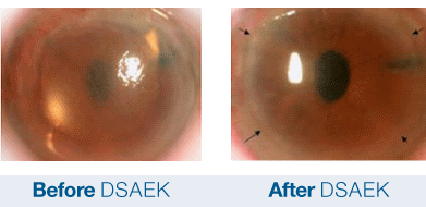
As the disease progresses, surgical intervention maybe necessary including a full-thickness corneal transplant or Descemet Stripping Automated Endothelial Keratoplasty (DSAEK). DSAEK is a partial thickness cornea transplant procedure that involves selective removal of the patient’s Descemet membrane and endothelium, followed by transplantation of donor corneal endothelium in addition to donor corneal stroma. This transplanted tissue is approximately 100-200 microns thick.
The Fraser Eye Care Center Doctors have either authored or reviewed and approved this content.